 Naru News日本語
Home › Glossary › Everyday science
What is the glymphatic system, thought to wash the brain during sleep?
Also called: Glymphatic pathway

In one minute
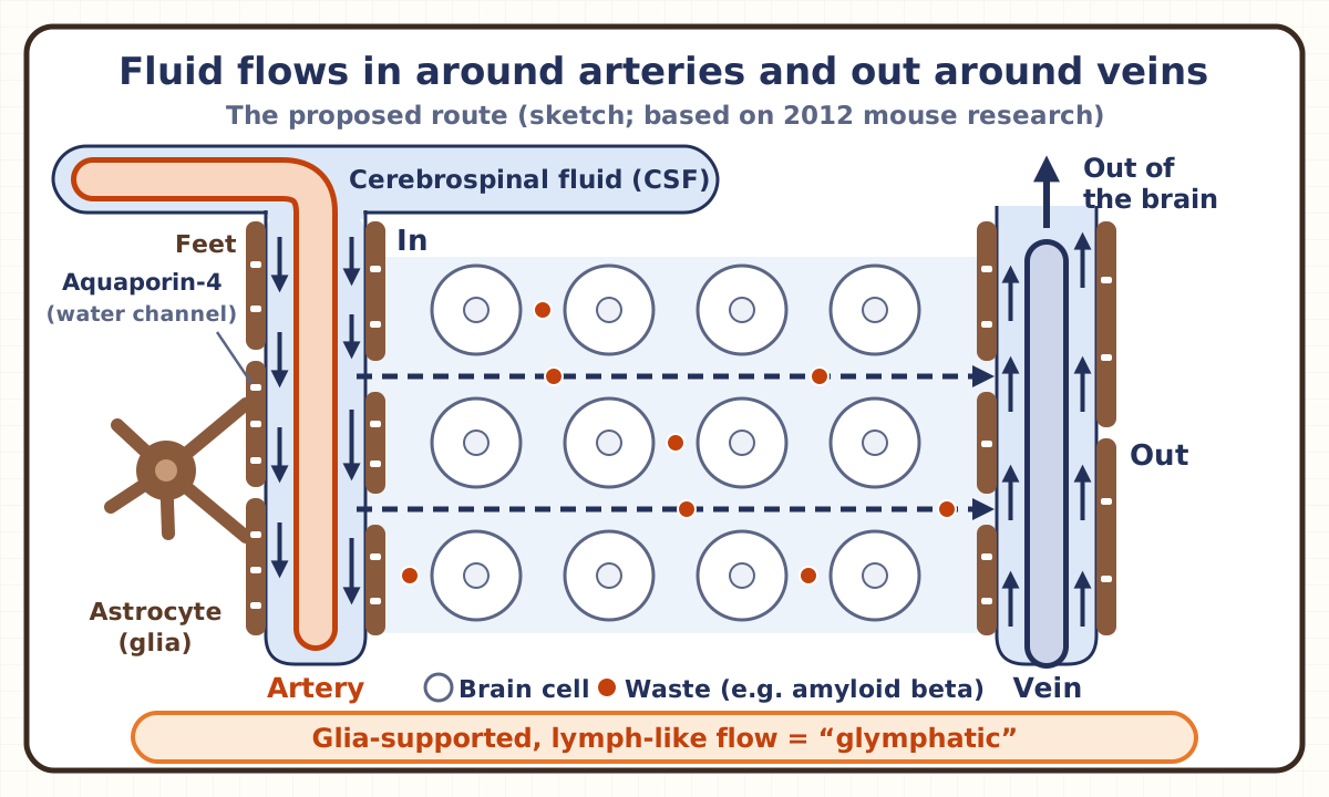
The glymphatic system is a proposed system in which the fluid around the brain and spinal cord (the bundle of nerves inside the backbone), called cerebrospinal fluid, seeps into the brain through narrow spaces around blood vessels, washes away waste that builds up around cells (the leftovers of cells’ work), and carries it out.
Elsewhere in the body, lymph vessels (thin tubes separate from blood vessels) collect leftover fluid and waste from around cells, but there are none inside the brain. In 2012, a flow of fluid that stands in for them was found in mouse brains. Because this lymph-like flow is supported by glia (cells that support nerve cells), it was named by combining “glia” and “lymphatic.”
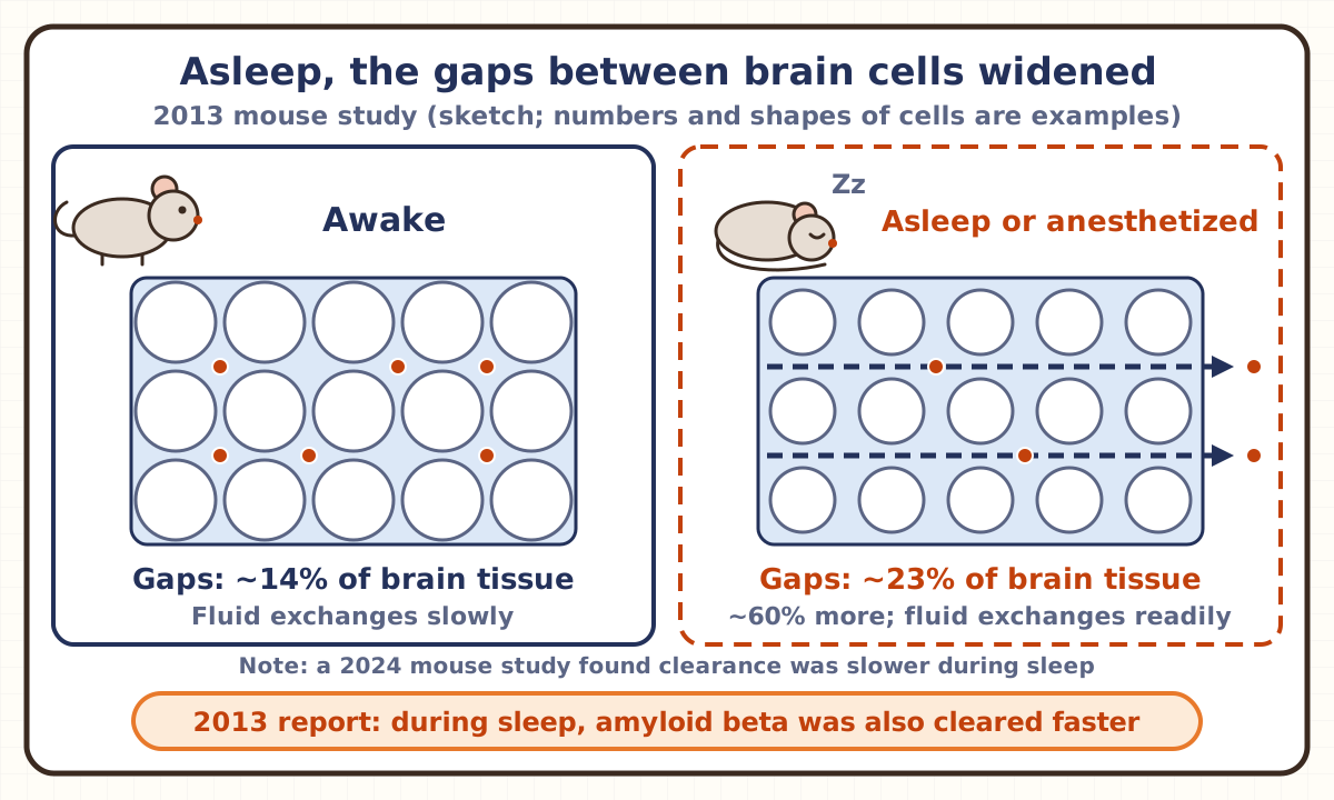
The next year, the spaces between brain cells were reported to widen during sleep in mice, with waste cleared faster, and the idea that “sleep cleans the brain” spread widely. In people, too, cerebrospinal fluid has been seen moving in and out in big waves during sleep. But a mouse study found clearance is actually slower during sleep, so whether sleep really speeds up the cleaning is still debated.
A common mix-up
“It has already been proven that sleep washes the brain.”
Half right. In a 2013 mouse study, during sleep or under anesthesia, the share of brain tissue taken up by the spaces between cells rose from about 14% when awake to about 23%, an increase of about 60%, and amyloid beta (fragments of protein that build up in the brains of people with Alzheimer’s disease) was cleared faster. In people, too, MRI has shown cerebrospinal fluid moving in and out in big waves during sleep. But a 2024 mouse study that measured in a different way found that material was actually cleared from the brain more slowly during sleep and anesthesia. And it has not yet been shown how much waste the fluid waves seen in people actually carry out. “Sleep washes the brain” is a leading idea, but it cannot yet be called an established fact.
Explained in pictures
You’re free to use these figures in lessons, slides and posts if you credit Naru News (naru.news). CC BY 4.0.
Think of it like this
Washing a shopping street at night
A shopping street is crowded during the day and hard to clean. So imagine that at night, when the shops have closed and the people have gone, water is let in from a channel at one end of the street and pushes the litter on the road all the way to a drain at the other end.
The glymphatic system is thought to work in the same way. The channel that brings in the water is the space around arteries. The littered street is the space between brain cells, and the drain is the space around veins. The water is cerebrospinal fluid, and the litter is waste such as amyloid beta. In mice, the spaces between cells were reported to widen during sleep, so water passes more easily, like an empty street at night.
But the comparison has limits. On a street you can see the water flow, but between brain cells it is still debated whether the fluid itself moves like a river, carrying material along, or whether the fluid barely moves and material just slowly spreads out, like a drop of ink in a glass of water. Whether cleaning really goes faster during sleep is also in question, since a study found the opposite. And nobody in the brain pours the water. What pushes the fluid is thought to be blood vessels pulsing with each heartbeat, and vessels slowly widening and narrowing.
Key points
Doesn’t the brain have lymph vessels?
Elsewhere in the body, lymph vessels collect the fluid that seeps out and is left over around cells, along with unneeded material in it, and carry it away. Lymph vessels are thin tubes separate from blood vessels, and the fluid inside them is called lymph. Inside brain tissue, however, there are no lymph vessels. So it was thought that the brain must clear away material around its cells in some other way.
The clue was cerebrospinal fluid, the fluid that fills the space around the brain and spinal cord and the spaces inside the brain. A 2012 mouse study showed that a large share of the cerebrospinal fluid around the brain enters brain tissue through the spaces around blood vessels and circulates between the cells.
Then where do the fluid and waste go after passing between the cells? In 2015, two research groups reported, one after the other, that the tough membrane wrapping the mouse brain has lymph vessels. These vessels carried cerebrospinal fluid and material in it, and connected to lymph nodes deep in the neck (small bean-shaped organs that lymph vessels lead to). One group showed that these vessels also take up fluid that has left the spaces between brain cells by way of the glymphatic system. There are no lymph vessels inside brain tissue, but there are some in the membrane around the brain.
Why is it called “glymphatic”?
The name joins “glia” and “lymphatic.” Glia is the collective name for the cells in the brain that support nerve cells. One kind, the astrocyte, wraps the brain’s blood vessels with the ends of its arm-like branches (its “feet”). The narrow gap between a vessel’s wall and these feet is the space around the blood vessel. Aquaporin-4, a protein that forms a passage for water, is concentrated in those feet.
The 2012 study watched the brains of living mice with a microscope that can see a little below the brain’s surface. It caught small glowing markers entering with cerebrospinal fluid through the spaces around arteries that go into the brain, and leaving along routes around veins. In mice whose astrocytes lacked aquaporin-4, the fluid flowed in more slowly, and the clearance of material between cells fell by about 70% compared with normal mice. Amyloid beta, thought to be involved in Alzheimer’s disease, was also cleared less well.
Because this flow carries waste like the body’s lymph and is supported by astrocytes, a kind of glia, the research group named it the “glymphatic system,” a word made from glial and lymphatic.
Why is it linked to sleep?
In 2013, the same research group compared mouse brains when the mice were awake with when they were asleep or under anesthesia. The spaces between brain cells took up about 14% of brain tissue when awake, but about 23% during sleep and anesthesia, an increase of about 60%. With wider spaces, cerebrospinal fluid got in between the cells more easily and swapped places with the fluid already there much more actively. And the rate at which amyloid beta was cleared from the brain also rose during sleep.
There was a reason to test anesthesia too. The sleeping mice were measured at midday and the awake mice in the evening, so the difference might have come from the time of day. So the researchers gave anesthesia to awake mice in the evening and measured again, and the spaces widened in the same way in the same mice. They concluded that the spaces change with whether the mice are awake, not with the time of day.
The paper suggested that sleep may restore us because, during sleep, the brain can better remove potentially harmful waste that builds up while we are awake. The result came to be widely described as “sleep cleans the brain.”
So what pushes the fluid during sleep? A 2025 mouse study found that during non-REM sleep (sleep without rapid eye movements, which includes deep sleep; sleep with rapid eye movements and more dreaming is REM sleep), the level of a brain substance involved in alertness and attention (norepinephrine) slowly rises and falls, and blood vessels widen and narrow along with it. When the researchers set off this slow vessel movement themselves, more cerebrospinal fluid flowed into the brain, so they think the vessel movement acts like a pump that pushes fluid in. In mice given a sleep medicine, both the rise and fall of this substance and the fluid flow became weaker. These are all results in mice, and it is not yet known whether the same happens in people.
Has it been confirmed in people?
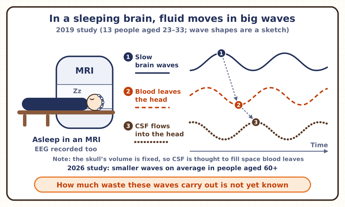
In people, you can’t look inside the head with a microscope as in mice. A 2019 study used fast MRI and EEG (brain-wave recording) together to observe 13 people aged 23 to 33 while they slept inside an MRI scanner. During non-REM sleep, a slow, large brain wave was followed by blood leaving the head, which was followed by cerebrospinal fluid flowing into the head, and this sequence repeated. Because the space inside the skull is fixed, cerebrospinal fluid is thought to flow in to fill the room left as blood leaves. In the sleeping brain, cerebrospinal fluid was moving in and out in waves big enough to see on MRI. But which change causes which is still being worked out.
In 2021, a study compared people who had been given a substance that shows up on MRI in their cerebrospinal fluid: 7 who stayed awake all night and 17 who slept as usual. In those who stayed awake, the agent was slower to leave many parts of the brain the next morning. Sleeping the following night did not make up for the delay.
In 2026, a study had 31 people aged 18 to 39 and 41 people aged 60 or over sleep in an MRI scanner late at night. In the young people, cerebrospinal fluid waves grew bigger during sleep, but in those aged 60 or over they grew far less on average, and they stayed smaller at every stage of non-REM sleep. While the participants were awake, the researchers also tested how well their brain’s blood vessels respond. Holding your breath raises the carbon dioxide in your blood, which normally increases blood flow in the brain. This response, and the rise in blood flow when the brain works, were weaker in those aged 60 or over. The researchers say that weaker slow brain waves in deep sleep and sluggish blood-vessel responses both played a part in the smaller waves, but could not explain all of it, so other factors are likely involved. Some people aged 60 or over had waves as large as young people’s.
The same year, the same lab reported an experiment in which, while 14 healthy people slept, the researchers watched their brain waves and played a quiet 0.05-second burst of noise (pink noise, a kind of hiss) timed to the peaks of the slow waves. The sound was too quiet to wake them, and both the slow brain waves and the cerebrospinal fluid waves became larger. But it has not yet been shown whether more waste was carried out, or whether it is good for sleep, memory or health.
What is the opposite result?
In 2024, another research group studied mice with a different method and reported the opposite result. They put a small glowing dye deep in the brain and kept measuring its brightness with a thin optical fiber at the front of the brain, some distance away. How fast the dye spread out between cells did not change whether the mice were awake, asleep or under anesthesia. And the dye was cleared from the brain clearly more slowly during sleep and anesthesia than when awake.
The paper says this is the opposite of the idea that clearance speeds up during sleep. It suggests that when dye seemed to get further into the brain during sleep in earlier studies, this may not have been because fluid flowed in better, but because it left more slowly. Think of the water level in a bathtub: it rises just the same whether you let more water in or narrow the drain so less goes out. The paper also says that larger substances may behave differently.
There is disagreement on a more basic point too: whether the fluid between brain cells moves along like a river, carrying material with it, or material just slowly spreads out, like ink dropped in a glass of water. A 2022 paper written with the researchers who first described the system also says that how much bulk flow there is between cells is still an open question. Including the link to sleep, which view is right has not been settled.
Is it linked to disease?
Amyloid beta is a small fragment of protein made in the brain, and in the brains of people with Alzheimer’s disease it clumps and builds up. The 2012 study suggested that the glymphatic system may remove amyloid beta from the brain, and that a weaker flow might be one reason proteins build up. In 2022, a Japanese research group also reported that in mice, tau, another protein linked to dementia, moves through this system from the brain into the cerebrospinal fluid and leaves the brain through lymph nodes in the neck.
But this is still at the stage of an idea. In mice, weakening this flow has been shown to make amyloid beta harder to clear, but it has not been confirmed that in people, a weaker system causes Alzheimer’s disease. The 2026 study finding smaller fluid waves with age did not include anyone diagnosed with such a disease, and the researchers hope to follow some of the participants to see whether smaller waves are linked to the risk of disease.
So it cannot yet be said, based on this system, that doing something in particular will prevent dementia.
So does sleep clean the brain or not?
Here is what is known so far. In mice, cerebrospinal fluid enters the brain through the spaces around blood vessels, passes between cells and flows out, and when this flow is weakened, amyloid beta and other material are cleared less well. In people, cerebrospinal fluid has been seen moving in and out in big waves during sleep, and after a night without sleep, a substance was slower to leave the brain.
What is still debated is whether sleep makes this flow and clearance faster or slower. Even in mice, one study found faster clearance during sleep and another found slower clearance. And it has not yet been shown how much waste the fluid waves seen in people actually carry out.
In short, fluid moving a lot in the brain during sleep has been seen in people too. But it cannot yet be said for certain that sleep cleans the brain better.
Check yourself
Q1A 2013 mouse study compared brains when the mice were awake with when they were asleep or under anesthesia. During sleep and anesthesia, what had happened to the spaces between brain cells?
Answer and explanation
Answer: B They were wider than when awake
During sleep and anesthesia, the share of brain tissue taken up by the spaces between cells rose from about 14% when awake to about 23%, about 60% more. With wider spaces, cerebrospinal fluid got in between the cells more easily and the fluid exchanged well, and amyloid beta was cleared faster. However, another mouse study in 2024 found the opposite: material was cleared from the brain more slowly during sleep.
Q2A 2019 study examined people’s brains during sleep with MRI and brain-wave recording at the same time. During non-REM sleep (sleep without rapid eye movements), what was happening just before cerebrospinal fluid flowed into the head?
Answer and explanation
Answer: C Blood was flowing out of the head
Just before cerebrospinal fluid flowed in, blood was leaving the head, and before that came a slow, large brain wave. Because the space inside the skull is fixed, the fluid is thought to flow in to fill the room left as blood leaves. Blood was not flowing into the head, and its amount was not staying the same: it was flowing out. But which change causes which is still being worked out.
Q3A 2024 mouse study put a glowing dye deep in the brain and compared how fast it was cleared from the brain when the mice were awake, asleep or under anesthesia. How fast was the dye cleared during sleep and anesthesia?
Answer and explanation
Answer: A Slower than when awake
The dye was cleared from the brain clearly more slowly during sleep and anesthesia than when awake. How fast the dye spread out between cells did not change in any state. This is the opposite of the earlier idea that clearance speeds up during sleep, and researchers are still debating how sleep and brain cleaning are related.
Related terms
- Cerebrospinal fluid
- Amyloid beta
- Alzheimer’s disease
- Non-REM sleep
- Glial cell
- Astrocyte
- Aquaporin-4
- Lymphatic vessel
- Meningeal lymphatic vessels
- Aging
- Epigenetic clock
Sources and how we checked
- Iliff et al., “A paravascular pathway facilitates CSF flow through the brain parenchyma and the clearance of interstitial solutes, including amyloid β” (University of Rochester and others; Science Translational Medicine 4(147), 147ra111, August 2012; abstract [the brain lacks a lymphatic circulation, a large share of subarachnoid CSF cycles through the brain interstitial space, in vivo two-photon imaging in mice, influx along spaces around penetrating arteries and clearance along paravenous routes, slowed influx and a ~70% reduction in interstitial solute clearance in mice lacking aquaporin-4, suppressed clearance of amyloid β, and possible protein mis-accumulation when the pathway is impaired]; record on Europe PMC, read November 17, 2026)
- University of Rochester Medical Center, “Scientists Discover Previously Unknown Cleansing System in Brain” (August 15, 2012; why it was named glymphatic [it acts much like the lymphatic system but is managed by glial cells], astrocyte end feet and water channels, and two-photon microscopy in living mice; read November 17, 2026)
- Xie et al., “Sleep drives metabolite clearance from the adult brain” (University of Rochester and others; Science 342(6156), pp. 373–377, 2013; abstract [in live mice, natural sleep or anesthesia is associated with a 60% increase in the interstitial space, a striking increase in exchange between CSF and interstitial fluid, a faster rate of β-amyloid clearance during sleep, and the suggestion that sleep’s restorative function may come from removing waste] and main text [an interstitial volume fraction of about 14% awake versus about 23% in sleep or anesthesia, and that sleep was measured at midday and wakefulness in the evening, with anesthesia in the same mice in the evening also widening the space, so the sleep-wake state rather than time of day was proposed as the cause]; record on Europe PMC and full text on PMC [PMC3880190], read November 17, 2026)
- Fultz et al., “Coupled electrophysiological, hemodynamic, and cerebrospinal fluid oscillations in human sleep” (Boston University and others; Science 366(6465), pp. 628–631, November 2019; abstract [accelerated neuroimaging, in non-REM sleep slow neural waves followed by hemodynamic oscillations coupled to CSF flow, and macroscopic waves of CSF flow in the sleeping brain] and main text [due to constant intracranial volume, more CSF flows into the head when less volume is occupied by blood, and causal links still to be tested in animals]; record on Europe PMC and full text on PMC [PMC7309589], read November 17, 2026)
- Boston University College of Engineering, “Are We ‘Brain Washed’ during Sleep?” (November 1, 2019; the 13 participants aged 23 to 33 in the 2019 study, sleeping in an MRI scanner wearing EEG caps, the order of neural change, blood flowing out of the head and CSF flowing in, and causal links still being open; read November 17, 2026)
- Eide et al., “Sleep deprivation impairs molecular clearance from the human brain” (University of Oslo and others; Brain 144, pp. 863–874, 2021; abstract [the contrast agent gadobutrol given intrathecally, 7 people with one night of total sleep deprivation compared with 17 who slept, impaired clearance from most brain regions the next morning, not made up by sleep the following night]; record on Europe PMC, read November 17, 2026)
- Miao et al., “Brain clearance is reduced during sleep and anesthesia” (Imperial College London and others; Nature Neuroscience 27(6), pp. 1046–1050, 2024; abstract and main text [male mice, dye injected into the caudate putamen and measured in the frontal cortex with an optical fiber, diffusion unchanged across wake, sleep and anesthesia, clearance markedly reduced during sleep and anesthesia, the opposite conclusion of earlier work, and that larger molecules may behave differently]; full text on Europe PMC [PMC11156584], read November 17, 2026)
- Hauglund et al., “Norepinephrine-mediated slow vasomotion drives glymphatic clearance during sleep” (University of Copenhagen, University of Rochester and others; Cell 188(3), pp. 606–622, February 2025, online January 8, 2025; abstract [in mouse non-REM sleep, synchronized slow oscillations in norepinephrine, cerebral blood volume and CSF as the strongest predictors of clearance, stimulated arterial oscillations increasing CSF inflow so that vasomotion acts as a pump, and the sleep aid zolpidem suppressing norepinephrine oscillations and glymphatic flow]; record on Europe PMC, read November 17, 2026)
- Bailes et al., “The aging human brain exhibits reduced hemodynamic and cerebrospinal fluid flow waves during sleep” (Massachusetts Institute of Technology and others; Nature Aging, November 13, 2026; abstract and Results [31 adults aged 18 to 39 and 41 aged 60 or over, average rise in CSF power from wake to non-REM sleep of 3.82 dB in young and 1.15 dB in older adults, smaller CSF flow in older adults at each stage of non-REM sleep, smaller blood-flow responses after breath-holding and in a visual task, both neural and cerebrovascular changes contributing, an age effect remaining after accounting for them, and the link to frontal gray matter volume] and Discussion [some older adults resembled young adults, additional mechanisms probably contributing, and some age-related factors were not measured]; full text on nature.com, read November 17, 2026)
- MIT, “Waves that clear waste from the brain during sleep are impaired in older adults” (MIT News, November 13, 2026; the two age groups, sessions of about two hours starting late at night, that holding one’s breath raises carbon dioxide in the blood, which normally increases cerebral blood flow, that the decline is not just a result of less sleep, two vascular factors explaining much but not all of the decline, older adults with youthful waves, no one with a diagnosed neurodegenerative disorder, and plans to follow some participants; read November 17, 2026)
- MIT, “A burst of ‘pink noise’ may lead to more restorative sleep” (MIT News, September 9, 2026; 14 healthy volunteers, a 50-millisecond burst of pink noise timed to the peak of slow waves, too quiet to wake a sleeper, and larger slow waves and CSF waves. The paper is Levitt et al., Science Translational Medicine 18(866), 2026; its full text could not be read. Read November 17, 2026)
- Louveau et al., “Structural and functional features of central nervous system lymphatic vessels” (University of Virginia; Nature 523(7560), pp. 337–341, July 2015; abstract [the long-held view that the central nervous system lacks a classical lymphatic drainage system, functional lymphatic vessels lining the dural sinuses, carrying fluid and immune cells from the CSF, and connection to the deep cervical lymph nodes]; record on Europe PMC, read November 17, 2026)
- Aspelund et al., “A dural lymphatic vascular system that drains brain interstitial fluid and macromolecules” (University of Helsinki; Journal of Experimental Medicine 212(7), pp. 991–999, June 2015; abstract [lymphatic vessels in the mouse dura that absorb CSF from the subarachnoid space and brain interstitial fluid via the glymphatic system, and carry it to the deep cervical lymph nodes]; record on Europe PMC, read November 17, 2026)
- Bohr et al., “The glymphatic system: Current understanding and modeling” (a review with Iliff, Nedergaard and others as co-authors; iScience 25(9), 104987, 2022; main text [the name based on astroglial aquaporin-4, aquaporin-4 concentrated in the end feet wrapping blood vessels, end feet separating the perivascular spaces from the tissue with fluid able to pass through the gaps between them, the absence of lymphatic vessels in neural tissue, arterial pulsation as a driver, and that whether bulk flow is appreciable between cells remains an open question]; full text on Europe PMC [PMC9460186], read November 17, 2026)
- Shindo, “Cerebral small vessel disease and Alzheimer’s disease: the glymphatic system as an amyloid β clearance system” (in Japanese; Mie University; Neurological Therapeutics 36(4), pp. 524–527, 2019; for the Japanese name “gurinpatikku-kei”; read on J-STAGE on November 17, 2026)
- University of Tokyo, Japan Science and Technology Agency (JST) and Japan Agency for Medical Research and Development (AMED), press release on how tau protein is cleared from the brain (in Japanese; February 26, 2022; for the Japanese names “glia-lymph system” and “glymphatic system,” and that in mice tau moves through this system into the CSF and leaves the brain through lymph nodes in the neck; read November 17, 2026)
AI wrote this page from the sources above, and a different AI checked it against the text of those sources (November 17, 2026). A person with a master’s degree in science and a Japanese high-school science teaching license also read and checked it before publishing. Numbers are as of that date. If anything was hard to follow, you spot a mistake, or there’s a term you’d like explained, please write to [email protected].
Everyday science terms
- Dark oxygen
- Epigenetic clock
- You are hereGlymphatic system
- Human embryo model
- iPS cell
- Manganese nodule
- Moore’s law
- Stem cell